The X-Rays That Confirm Rottweiler Osteosarcoma
Seeing your beloved Rottweiler suddenly develop a limp is one of the most heart-wrenching and concerning experiences any dog owner can face. It's a moment that instantly triggers a cascade of worry, especially when you know that a breed as magnificent and powerful as a Rottweiler is unfortunately prone to certain serious health conditions. This guide aims to walk you through the critical role X-rays play in investigating that troubling limp, specifically when the dreaded possibility of osteosarcoma looms, and what you can expect from this crucial diagnostic step.
The Alarming Limp That Needs Immediate Attention
A limp in a Rottweiler is never something to ignore or hope will "just get better." Their substantial size and muscular build mean that even minor discomfort can escalate quickly, and more importantly, some underlying issues are incredibly serious. When you notice your Rottweiler favoring a leg, holding it up, or showing any sign of lameness, your first call should always be to your veterinarian. This isn't just about pain; it's about finding answers as swiftly as possible, because early detection can make a world of difference.
When to Be Most Concerned About a Limp
While any limp warrants a vet visit, certain factors should heighten your concern. If your Rottweiler is over the age of six, the risk of conditions like osteosarcoma significantly increases. However, it's not unheard of in younger dogs, so age shouldn't be the only factor in your decision to seek care. A sudden, severe limp, especially if there was no obvious injury, or if the limp progresses rapidly, making your dog reluctant to bear weight, is a huge red flag. Equally concerning is a persistent, low-grade limp that doesn't resolve with rest, or one that seems to improve slightly only to worsen again. This kind of insidious onset can sometimes characterize bone cancer, which is why swift investigation is key.
Many owners initially assume a sprain or a minor muscle pull, which is a natural first thought, but given the breed's predisposition, it’s vital to consider all possibilities. For more information on identifying early signs, you might find our guide on The First Limp That Turns Out to Be Bone Cancer particularly helpful.
The Initial Vet Examination and What It Reveals
When you bring your Rottweiler in for a limp, your vet will perform a thorough physical examination. This typically involves observing your dog's gait, both walking and sometimes trotting, to assess the degree and nature of the lameness. They'll meticulously palpate the affected leg, feeling for swelling, heat, pain response, and any abnormalities in the bone or soft tissues. They’ll also check the range of motion in all joints, carefully flexing and extending limbs.
During this examination, your vet will be trying to pinpoint the exact location of the pain. Is it in a joint? Is it along the shaft of a long bone? Is it a soft tissue injury? For Rottweilers, particular attention will be paid to the long bones of the limbs – the radius, ulna, tibia, and femur – as these are common sites for osteosarcoma. Don't be surprised if your vet wants to check the other limbs too; sometimes, pain in one area can cause compensatory lameness in another. They might even feel the lymph nodes and abdomen, looking for any other signs of systemic illness or swelling.
Why X-Rays Are the Gold Standard for Osteosarcoma Suspicion
Once the vet has localized the pain to a specific limb or area of bone, X-rays become the next, most crucial diagnostic step. For a large, deep-chested breed like a Rottweiler, X-rays offer a non-invasive way to look inside the bones and joints, providing vital clues that a physical exam simply can't. While they aren't the definitive diagnostic tool for cancer without a biopsy, X-rays are exceptionally good at revealing changes in bone structure that are highly suggestive of osteosarcoma.
What Osteosarcoma Looks Like on an X-Ray
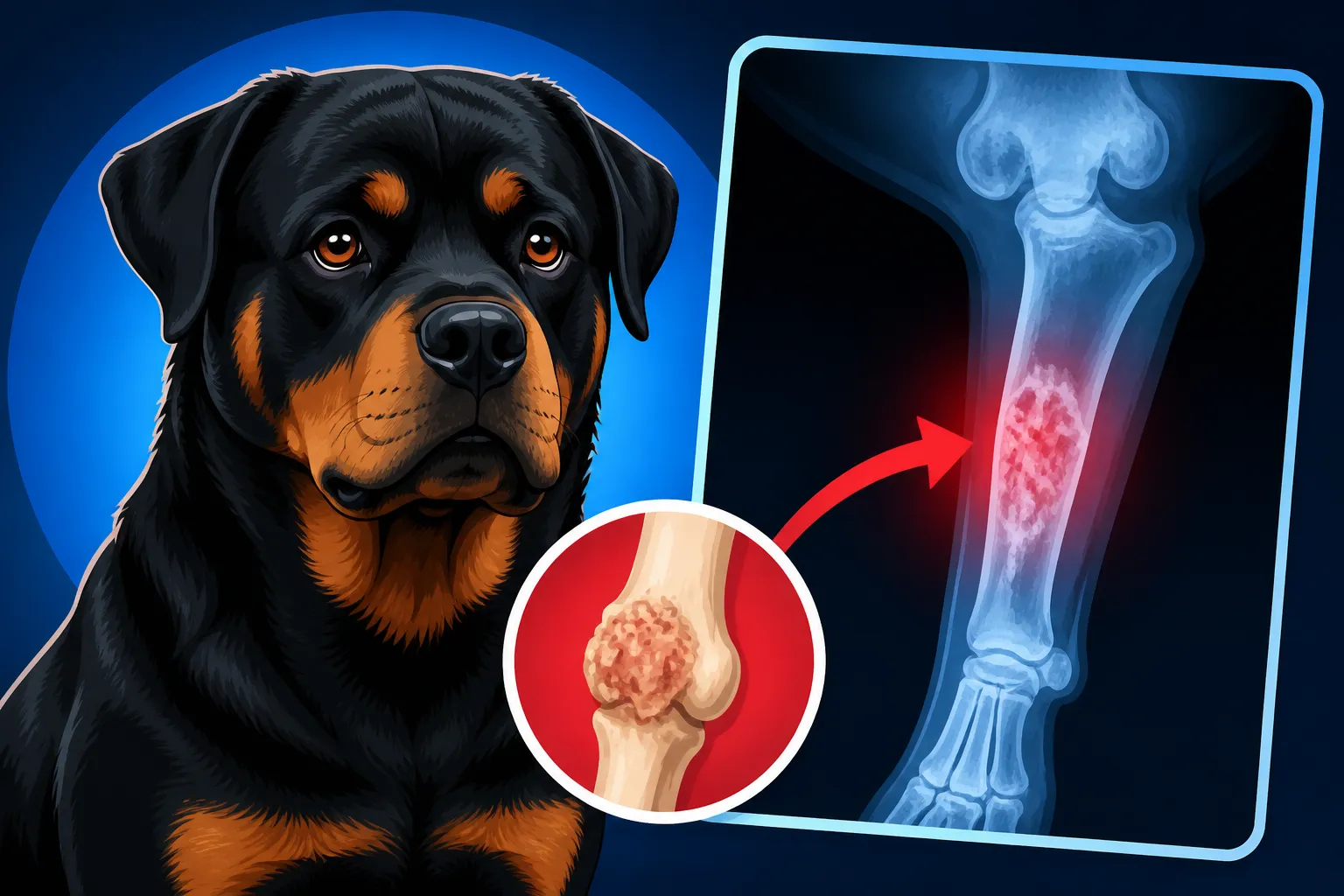
On a radiograph (X-ray image), normal bone appears as a solid, dense white structure. When osteosarcoma is present, however, the picture changes dramatically. The tumor essentially eats away at the healthy bone while also trying to produce abnormal bone tissue, leading to a very characteristic, often aggressive, appearance.
Vets and radiologists look for several key features:
- Lytic Lesions: These appear as dark, irregular areas on the X-ray, indicating bone destruction. The cancer cells are breaking down the normal bone architecture, creating "holes" or areas of decreased density.
- Blastic (or Sclerotic) Lesions: Conversely, some parts of the tumor may try to produce new, chaotic bone. These areas show up as irregular, increased densities or patches of white, often described as "fluffy" or disorganized.
- Periosteal Reaction: This is the body's response to irritation or inflammation of the periosteum, the outer membrane of the bone. With osteosarcoma, this reaction is often aggressive and irregular, sometimes appearing as a "sunburst" pattern where spicules of new bone radiate outwards, or a "moth-eaten" pattern indicating rapid, destructive growth.
- Cortical Destruction: The cortex is the hard outer layer of the bone. Osteosarcoma often breaks through this, making the bone look ill-defined or fractured.
- Soft Tissue Swelling: While X-rays are best for bone, they can also show generalized soft tissue swelling around the affected bone, which is common with aggressive tumors.
It's these distinct patterns – the mix of bone destruction and chaotic bone production, often with a dramatic periosteal reaction – that make X-rays incredibly useful in strongly suggesting a diagnosis of osteosarcoma. However, it's important to remember that other conditions, such as severe bone infections (osteomyelitis) or even fungal bone disease, can sometimes mimic the appearance of bone cancer. This is why further steps are often necessary. Our guide on Where Osteosarcoma Usually Appears on a Rottweiler provides more details on typical locations.
Beyond the Initial X-Ray Views
Just taking one X-ray view of a limb often isn't enough. Your vet will likely request at least two, often three or more, different views (e.g., cranio-caudal, medio-lateral, oblique projections) of the affected area. This is because bone lesions can be subtle, and a change might only be visible from a certain angle. These multiple views ensure that no part of the bone is hidden or obscured, providing a comprehensive picture.
For optimal imaging, especially with a powerful and sometimes restless dog like a Rottweiler, sedation is often recommended, if not required. This ensures the dog remains perfectly still, allowing for clear, crisp images without motion blur. It also allows the veterinary team to position the limb precisely, which is crucial for diagnostic accuracy. Sedation also makes the process much less stressful and painful for your Rottweiler, particularly if the limb is already very sore.
It's a scary time, and the costs associated with these diagnostics can add up. Many proactive owners invest in pet insurance. Companies like Healthy Paws can offer a real safety net, helping to cover a significant portion of these unexpected diagnostic and treatment expenses, which can be considerable when facing something like bone cancer.
Understanding the X-Ray Report and Next Steps
Once the X-rays are taken, your vet, or sometimes a veterinary radiologist, will meticulously examine the images. The report will describe any abnormalities found, noting their location, size, and characteristics (e.g., lytic, blastic, periosteal reaction). They will then discuss these findings with you, explaining what they mean in the context of your Rottweiler's symptoms and history.
The Role of Biopsy for Confirmation
Even if the X-rays show changes that are "highly suggestive" or "classic" for osteosarcoma, an X-ray alone cannot definitively confirm cancer. This is a critical point that many owners grapple with. To get a 100% confirmed diagnosis, a biopsy is necessary. A biopsy involves taking a small tissue sample from the affected bone, which is then sent to a veterinary pathologist for microscopic examination.
There are a couple of common biopsy methods:
- Needle Biopsy: This is less invasive and can sometimes be done under heavy sedation. A specialized needle is used to extract small core samples from the suspicious bone lesion. While less invasive, it can sometimes yield inconclusive results if the sample doesn't contain enough cancerous cells, or if it only hits areas of reactive bone rather than the tumor itself.
- Incisional Biopsy: This is a more invasive surgical procedure performed under general anesthesia. A small incision is made, and a piece of bone tissue is surgically removed. This generally provides a larger, more representative sample, increasing the likelihood of a definitive diagnosis.
Your vet will discuss the pros and cons of each method based on the X-ray findings and your dog's overall health. A confirmed diagnosis from a biopsy is crucial because it guides all subsequent treatment decisions, from pain management to more aggressive therapies like amputation and chemotherapy. You can learn more about managing the disease in our article on Pain Control for a Rottweiler With Bone Cancer.
Further Imaging and Staging for Treatment Planning
If osteosarcoma is confirmed, or if the X-ray findings are so compelling that your vet recommends moving forward with treatment based on strong suspicion, further imaging will be needed for "staging." Staging refers to determining if the cancer has spread to other parts of the body. Unfortunately, osteosarcoma is highly metastatic, meaning it tends to spread rapidly, often to the lungs.
- Chest X-rays: These are almost always performed to check for lung metastasis. Multiple views of the chest are taken to increase the chances of detecting small tumors that might have spread. Finding visible lung metastases significantly changes the prognosis and treatment plan.
- CT or MRI Scans: While not always necessary for diagnosis, these advanced imaging techniques can be incredibly valuable, especially for surgical planning. A CT scan, for instance, provides highly detailed cross-sectional images of the bone and surrounding tissues, allowing surgeons to precisely assess the extent of the tumor and plan the safest and most effective surgical approach if amputation is being considered. These scans can also help detect subtle metastases that might be missed on standard X-rays.
This extensive diagnostic process can feel overwhelming, but each step provides essential information that helps your veterinary team create the most effective, compassionate plan for your Rottweiler. Having pet insurance from a provider like Healthy Paws can seriously ease the burden of these advanced diagnostics and subsequent treatments.
| X-Ray Feature | Normal Bone | Suspicious for Osteosarcoma |
|---|---|---|
| Bone Density | Uniform, solid white | Irregular mixed areas of darkness (lysis) and increased white (sclerosis) |
| Bone Margins | Smooth, well-defined | Blurry, indistinct, "moth-eaten" appearance |
| Periosteum | Not visible | Visible, irregular, often "sunburst" or "onion peel" reaction |
| Cortex | Intact, solid outer layer | Eroded, broken, or permeated by tumor |
| Soft Tissue | No significant swelling | Localized swelling around affected bone |
| Location | No specific predisposition for abnormalities | Often affects metaphysis (ends) of long bones (radius, tibia, femur) |
Financial Realities and Support for Rottweiler Owners
Let's be real: diagnosing and treating serious conditions like osteosarcoma in a large breed like a Rottweiler can be incredibly expensive. This is a stark reality that many owners face, often adding immense stress to an already emotional situation. It’s not just the initial X-rays; it's the biopsy, chest X-rays, potential CT scans, specialist consultations, and then the treatment itself, whether that involves surgery, chemotherapy, or palliative care.
Costs Associated with Diagnostic Imaging
The cost of X-rays can vary significantly based on your location (urban vs. rural), the veterinary clinic, and the number of views required. Generally, you might expect to pay anywhere from $200 to $500 for a set of diagnostic X-rays on a large dog. If sedation is needed, that will add to the bill. A bone biopsy, depending on the method and whether it requires referral to a specialist surgeon, could range from $500 to $1,500 or more. Advanced imaging like CT scans often run into thousands of dollars.
These are significant sums, and they come at a time when you're already grappling with the fear for your dog's health. This is precisely why preparedness is so important.
The Lifeline of Pet Insurance
This is where pet insurance can be a genuine game-changer. For a condition like osteosarcoma, which can lead to treatments such as amputation (see Amputation Recovery Week by Week for a Rottweiler) and subsequent Chemotherapy Protocols Used on Rottweilers, the lifetime costs can easily run into tens of thousands of dollars. Imagine having the ability to say "yes" to every recommended diagnostic test and treatment plan without having to agonize over the financial impact.
Many owners choose to enroll their Rottweiler in pet insurance when they are puppies, precisely to avoid situations where a condition like cancer is considered "pre-existing" and therefore not covered. Companies like Healthy Paws understand the significant health challenges specific breeds like Rottweilers face and offer comprehensive plans that can cover accidents, illnesses, surgeries, and even some diagnostic tests. Having a policy in place could mean that after your deductible, Healthy Paws could reimburse a substantial percentage of your vet bills, easing the financial strain immensely. This allows you to focus on your Rottweiler's care, rather than the escalating costs. You can explore options like Rottweiler Pet Insurance and the $204 Average to understand how these plans work.
The emotional toll of a cancer diagnosis is heavy enough. Knowing you have financial support to provide the best possible care for your Rottweiler can make a world of difference in a truly heartbreaking situation. While it doesn't remove the pain, it ensures you can make decisions based on what's best for your beloved companion.
Disclaimer
The information provided in this article is for educational and informational purposes only and does not constitute veterinary medical advice. Always consult with a qualified veterinarian for any health concerns or before making any decisions about your pet's health or treatment.