Mast Cell Tumors on a Rottweiler’s Skin
Finding a new lump or bump on your Rottweiler's skin can send a shiver down any owner's spine. Our big, sturdy companions are prone to various skin growths, and while many are benign, one of the more concerning possibilities is a mast cell tumor. It’s a scary thought, but understanding these tumors – how they look, how they're diagnosed, and what treatment involves – can make all the difference in navigating this challenging journey for your beloved dog.
What Exactly Are Mast Cell Tumors on a Rottweiler's Skin?
When you hear the term "mast cell tumor," it probably sounds serious, and often, it can be. These aren't just any old skin growths; they're a type of cancer that originates from mast cells, which are a normal and vital part of your Rottweiler's immune system. Think of mast cells as the body's little alarm bells, packed with histamine and other inflammatory chemicals. They're usually busy fighting off parasites, repairing tissues, and dealing with allergic reactions. The problem arises when these cells start to grow uncontrollably and form a tumor.
What makes mast cell tumors particularly tricky is their unpredictable nature. They can pop up anywhere on the body, but the skin is the most common site, accounting for about 50% of all cases. For a large breed like a Rottweiler, these can sometimes be hidden by their dense coat, making regular hands-on checks even more important. It's truly unnerving how varied they can be, making it impossible to tell just by looking whether a lump is harmless or potentially life-threatening. This is why any new lump on your Rottweiler always warrants a trip to the vet.
Understanding Mast Cells and Their Role
To really grasp what a mast cell tumor is, let's dive a little deeper into the cells themselves. Mast cells are a type of white blood cell, essentially first responders in your dog's immune system. They're found in various tissues throughout the body, particularly those exposed to the external environment, like the skin, respiratory tract, and gastrointestinal tract. When your Rottweiler encounters an allergen or a perceived threat, these cells degranulate, meaning they release their stored chemicals. This release causes local inflammation – think itching, swelling, and redness – all part of the body's defense mechanism.
In the case of a mast cell tumor, these cells become cancerous. They not only multiply excessively but can also continue to degranulate spontaneously, even without a trigger. This uncontrolled release of inflammatory chemicals can lead to a whole host of problems, not just localized at the tumor site. It's why a Rottweiler with a mast cell tumor might experience a fluctuating lump, or even more generalized symptoms if the tumor is particularly aggressive or has spread.
Why Rottweilers Are More Susceptible to Mast Cell Tumors
It’s an unfortunate reality that certain breeds, including our beloved Rottweilers, seem to have a higher predisposition to developing mast cell tumors. While the exact genetic mechanisms aren't fully understood, breed-specific tendencies are well-documented. Other breeds often cited include Boxers, Boston Terriers, Bulldogs, and Golden Retrievers. This breed-specific risk means that if you own a Rottweiler, you need to be particularly vigilant about checking for new skin growths and acting quickly if you find one.
The average age for diagnosis in dogs is typically around 8 to 10 years old, putting them right in their senior years, but these tumors can certainly appear in younger dogs as well. This predisposition doesn't mean every Rottweiler will get a mast cell tumor, but it does highlight the importance of proactive care and regular veterinary check-ups, especially as your dog approaches the age where Cancer Screening for a Rottweiler Past Six becomes even more critical.
Grades of Mast Cell Tumors and What They Mean
Once a mast cell tumor is definitively diagnosed, often through a biopsy, it will be "graded." This grading is absolutely crucial because it tells us how aggressive the tumor is likely to be and helps predict prognosis and guide treatment. There are generally two main grading systems you might hear about: the Patnaik system (Grade I, II, III) and the newer, often preferred, Kiupel system (Low Grade or High Grade).
Here's a simplified breakdown:
| Grade (Kiupel System) | Characteristics | Behavior | Prognosis |
|---|---|---|---|
| Low Grade | Cells look relatively normal, well-differentiated | Less aggressive, slower to spread, often cured with complete surgical removal. | Generally good with appropriate treatment; longer survival times are common. |
| High Grade | Cells look abnormal, poorly differentiated | Very aggressive, rapid growth, high likelihood of local recurrence and metastasis. | Guarded to poor, even with aggressive treatment; shorter survival times. |
A low-grade tumor, while still cancerous, is generally much easier to manage and has a better outlook. High-grade tumors, on the other hand, are a much more serious concern, requiring aggressive intervention and often a multi-modal approach to treatment. It's worth noting that even a visually innocent-looking bump could be a high-grade tumor, emphasizing why proper diagnostic testing is non-negotiable.
Recognizing the Signs and Symptoms of Mast Cell Tumors
One of the most frustrating things about mast cell tumors is how deceptive they can be. They don't always look like a classic "cancerous lump," which can make initial identification incredibly challenging for owners. You might expect something firm and immovable, but a mast cell tumor can appear in a dizzying array of forms, mimicking many other benign skin conditions. This is why the saying "never ignore a new lump" is particularly true for Rottweilers, given their increased risk.
It's common for owners to discover these lumps during routine petting or grooming sessions. They might feel like a small nodule, a raised area, or even just a subtle change in skin texture. Because of their varied appearance, many owners initially dismiss them as something innocuous, like a bug bite or a fatty lipoma. But for a Rottweiler, especially one past middle age, every lump should be viewed with a healthy dose of suspicion and promptly evaluated by a vet. This proactive approach can literally be a lifesaver.
What Mast Cell Tumors Look Like on the Skin
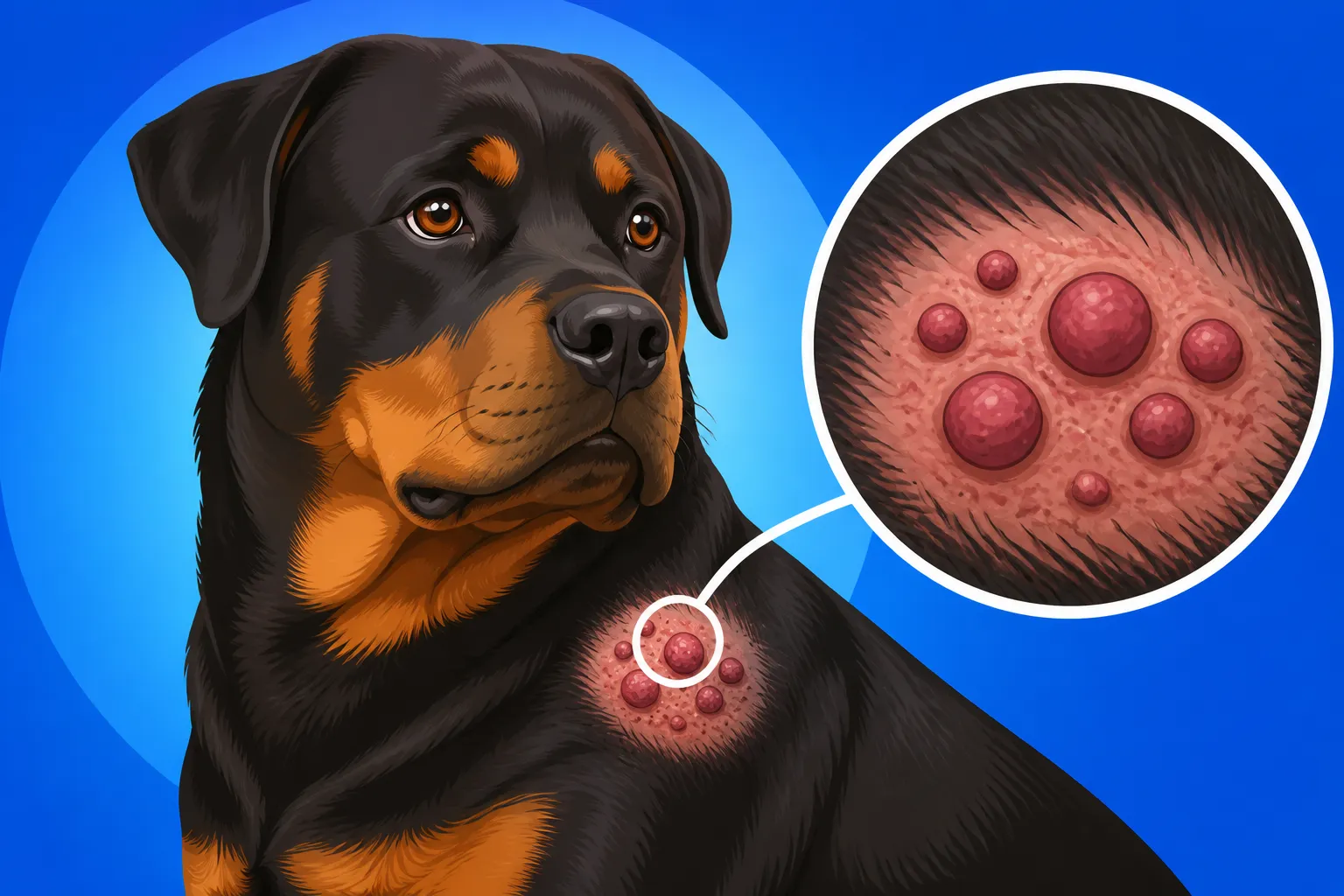
Mast cell tumors on the skin can be incredibly diverse in appearance, which is why relying solely on visual inspection is a gamble. They might present as:
- Single or Multiple Lumps: Sometimes you'll find just one, other times several.
- Varied Size: From tiny pea-sized bumps to large, sprawling masses.
- Location: While most commonly on the trunk and limbs, they can appear anywhere, including the head, neck, and paws.
- Texture: They can be soft and squishy, firm, or even rubbery. Some might feel like they're attached to underlying tissue, while others seem superficial.
- Color: Often pink or red, but can also be flesh-colored, black, or even ulcerated. An ulcerated tumor might have a scab or crust, mimicking a wound.
- Hair Loss: Sometimes the skin over the tumor may have thinning hair or be completely bald.
One common characteristic that can be a tell-tale sign is their tendency to wax and wane. This means the lump might appear to get bigger and redder one day, then shrink and become less noticeable the next. This fluctuation is often due to the mast cells within the tumor spontaneously degranulating, releasing histamine and causing localized swelling and inflammation, then subsiding. This "itchy bump syndrome" is a strong indicator that the lump could be a mast cell tumor, but it’s not always present. It's a key reason why any lump that changes in size or appearance needs immediate attention. If you've found Lumps on a Rottweiler That Need Testing, your vet will likely start with a fine needle aspirate.
Beyond Skin Lesions: Other Warning Signs
While most mast cell tumors are found on or just under the skin, it’s important to remember they can also develop internally, affecting organs like the spleen, liver, lymph nodes, and bone marrow. These internal tumors are often more aggressive and much harder to detect early because their symptoms are more vague and non-specific.
If your Rottweiler has an internal mast cell tumor, you might notice signs such as:
- Gastrointestinal issues: Vomiting, diarrhea, loss of appetite, black tarry stools (melena) which indicates digested blood. This is because the histamine released by mast cells can stimulate stomach acid production, leading to ulcers.
- Lethargy or weakness: Your dog might seem unusually tired or less energetic.
- Weight loss: Unexplained loss of condition despite a normal appetite.
- Abdominal pain or swelling: If the spleen or liver is affected, your dog might be uncomfortable when touched or show signs of a distended belly.
- Bleeding problems: In rare cases, massive histamine release can interfere with blood clotting.
These systemic signs can be concerning, and unfortunately, by the time they become obvious, the disease may be quite advanced. This again underscores the critical importance of regular vet check-ups and reporting any subtle changes in your Rottweiler’s behavior or physical condition. Being aware of Rottweiler Cancers Beyond the Bone helps owners remain vigilant for a range of symptoms.
The Darier's Sign: A Specific Indication
While not always present, a phenomenon known as "Darier's sign" can sometimes be observed with mast cell tumors. This occurs when you rub or scratch the tumor, and it rapidly becomes red, swollen, and itchy. This reaction is a direct result of the pressure causing the mast cells within the tumor to degranulate and release their inflammatory contents, primarily histamine.
If you gently palpate a lump on your Rottweiler and notice it quickly reddens, swells, or even causes your dog to react as if it's suddenly itchy, that's a very strong indicator that you're dealing with a mast cell tumor. While it's not a definitive diagnostic tool, it should certainly heighten your suspicion and accelerate your trip to the veterinarian for a proper diagnosis.
Diagnosis and Staging of Mast Cell Tumors in Rottweilers
The moment you discover a suspicious lump on your Rottweiler, the clock starts ticking. Early and accurate diagnosis is paramount for mast cell tumors, as their behavior can range from relatively benign to highly aggressive. It's a time filled with anxiety, but your veterinarian has a clear protocol to follow, designed to get you answers as quickly and definitively as possible. Don't hesitate or try to "watch it" for a few weeks; that time could be critical, especially for a fast-growing, high-grade tumor.
The diagnostic process usually involves several steps, starting with a simple, non-invasive test and potentially moving to more involved procedures depending on the initial findings. Your vet’s goal isn’t just to identify the lump, but to understand its full scope – its grade and whether it has spread.
Initial Vet Examination and Fine Needle Aspirate
The first thing your veterinarian will do is a thorough physical examination. They’ll carefully feel the lump, noting its size, location, texture, and whether it's fixed or movable. They'll also check surrounding lymph nodes, as these are common sites for metastasis (spread) in mast cell tumors.
The next, and most crucial, immediate step is usually a Fine Needle Aspirate (FNA). This involves using a small, thin needle to collect a sample of cells directly from the lump. The procedure is quick, generally well-tolerated by Rottweilers, and often doesn't even require sedation. The collected cells are then smeared onto a slide and examined under a microscope. Mast cells have a distinctive appearance, often containing dark granules, which allows skilled veterinarians and veterinary cytologists to often make a presumptive diagnosis of a mast cell tumor right in the clinic or with a quick lab turnaround.
An FNA is incredibly useful for providing a rapid initial answer, but it does have limitations. It can often confirm the presence of mast cells, but it cannot definitively determine the tumor's grade (low vs. high) or how deeply it has invaded tissue. For that, a more comprehensive sample is needed.
Biopsy and Histological Grading: The Definitive Diagnosis
If the FNA suggests a mast cell tumor, or if the results are inconclusive, the next step is a biopsy. A biopsy involves surgically removing a piece of the tumor (incisional biopsy) or, ideally, the entire tumor with a wide margin (excisional biopsy) and sending it to a specialized veterinary pathologist. This is the only way to get a definitive diagnosis and, critically, to determine the tumor's grade.
The pathologist examines the tissue under a microscope, looking at various cellular characteristics to assign a grade (using either the Patnaik or Kiupel system, as discussed earlier). This histological grading is the most important prognostic indicator for mast cell tumors. It tells your vet whether the tumor is likely to be slow-growing and localized, or aggressive and prone to spreading. The pathologist will also assess the "margins" – the healthy tissue surrounding the tumor that was removed. "Clean margins" mean all the cancerous cells appear to have been removed, which is always the goal. "Dirty" or "incomplete margins" mean some cancerous cells were left behind, which significantly increases the risk of recurrence and may necessitate further surgery or adjunctive therapies.
Staging the Disease: Beyond the Skin
Even if a mast cell tumor appears confined to the skin, it’s vital to determine if it has spread to other parts of the body. This process is called "staging," and it helps your vet understand the full extent of the disease and plan the most effective treatment strategy. Staging tests are particularly important for high-grade tumors or those in concerning locations.
Common staging procedures include:
- Regional Lymph Node Aspirate: The lymph nodes closest to the tumor are often the first place mast cell tumors spread. Your vet may aspirate these nodes to check for cancerous cells.
- Abdominal Ultrasound: This allows visualization of internal organs like the spleen and liver, which are common sites for metastasis. If abnormalities are seen, biopsies or aspirates may be taken.
- Chest X-rays: While mast cell tumors rarely spread directly to the lungs, chest X-rays can help assess overall lung health and rule out other issues.
- Bloodwork and Urinalysis: These tests assess your Rottweiler's general health, organ function, and can reveal signs of systemic inflammation or internal degranulation from the tumor.
Sometimes, more advanced imaging like CT scans or even bone marrow aspirates might be recommended for aggressive tumors or if systemic signs are present. The information gathered from staging helps your vet determine if your Rottweiler has localized disease or if it has unfortunately become a more widespread, systemic problem. This comprehensive approach is essential for giving your Rottweiler the best chance. When facing unexpected vet bills for diagnosis and treatment, many owners find that having pet insurance, such as through a provider like Healthy Paws, can be an absolute lifesaver, helping to manage the significant financial burden.
Treatment Options and Prognosis for Rottweilers with Mast Cell Tumors
Receiving a diagnosis of a mast cell tumor is undoubtedly terrifying, but it's important to remember that there are many effective treatment options available, and the prognosis can vary widely depending on the tumor's grade and stage. Your veterinary oncology team will work with you to develop a personalized treatment plan aimed at giving your Rottweiler the best possible outcome and maintaining a good quality of life. The approach is often multi-modal, meaning it might involve a combination of therapies.
Navigating these choices can feel overwhelming, especially when faced with complex medical information and potential costs. This is where a candid discussion with your vet about expectations, potential side effects, and financial considerations becomes crucial. While we always hope for the best, understanding all the possibilities helps you make informed decisions for your cherished companion.
Surgical Removal: The Primary Treatment
For mast cell tumors on the skin, surgical removal is almost always the first and most important line of defense. The goal of surgery is to remove the entire tumor, along with a wide margin of healthy tissue around it. This "wide margin" is critical because mast cell tumors often have microscopic extensions that aren't visible to the naked eye. In some cases, especially for larger or more invasive tumors, the surgeon might need to remove a significant amount of surrounding tissue, which could involve complex reconstructive surgery, particularly if the tumor is located on a limb or the head.
If the pathologist confirms that the surgical margins are "clean" (meaning no cancer cells were found at the edges of the removed tissue), and the tumor is low-grade, surgery alone might be curative. However, if the margins are "dirty" (incomplete removal), or if the tumor is high-grade, further treatments are almost certainly recommended to prevent recurrence and spread. This could mean a second surgery to achieve wider margins, or a move to adjunctive therapies like radiation or chemotherapy.
Chemotherapy and Radiation: When They Come Into Play
When surgery alone isn't enough, or if the disease has spread, your vet will likely recommend adjunctive therapies.
- Radiation Therapy: This is often recommended for tumors that are incompletely removed by surgery or are in locations where wide surgical excision isn't possible (e.g., around the eyes or on the paw). Radiation therapy targets any remaining cancer cells in the localized area, aiming to prevent recurrence. It typically involves multiple treatments under anesthesia over several weeks. Radiation can be highly effective for local disease control.
- Chemotherapy: This is usually reserved for high-grade mast cell tumors, tumors that have spread to regional lymph nodes or internal organs, or tumors that are inoperable. Chemotherapy uses drugs to kill cancer cells throughout the body. While it won't typically cure metastatic mast cell disease, it can often slow its progression and improve quality of life. Common chemotherapy drugs used for mast cell tumors include prednisone (which can reduce inflammation and tumor size), vinblastine, and oral tyrosine kinase inhibitors like Palladia or Kinavet. The choice of drug depends on the tumor's characteristics and your dog's overall health. Chemotherapy protocols are carefully managed to minimize side effects, which are generally less severe in dogs than in humans.
Understanding the complexity and cost of these treatments is important. For a Rottweiler, these bills can quickly add up, and having strong pet insurance coverage from a company like Healthy Paws can significantly reduce the financial stress, allowing you to focus on your dog's care. Many policies will cover a large percentage of specialist visits, diagnostics, and treatments like chemotherapy and radiation. However, it's critical to note that What a Rottweiler Policy Will Not Cover often includes pre-existing conditions, so getting insurance early is always a good idea.
Managing Expectations and Prognosis
The prognosis for a Rottweiler with a mast cell tumor depends heavily on several factors:
- Tumor Grade: Low-grade tumors generally have a much better prognosis than high-grade tumors.
- Clinical Stage: Tumors that are localized to the skin have a better prognosis than those that have spread to lymph nodes or internal organs.
- Location: Tumors on the extremities (like paws) or those that are small and easily removed often have a better outcome than those on the trunk, head, or mucocutaneous junctions (e.g., lips).
- Completeness of Surgical Removal: Achieving clean surgical margins is a strong positive prognostic indicator.
For low-grade, completely removed skin mast cell tumors, the prognosis can be excellent, with many Rottweilers living for many years post-surgery with no recurrence. For high-grade or metastatic tumors, the prognosis is more guarded, but even in these cases, chemotherapy or radiation can often extend life and maintain good quality for months or even a year or more.
It’s a tough discussion, but your vet will give you an honest assessment based on all diagnostic findings. They'll also discuss Palliative Care for a Rottweiler With Cancer options if curative treatment isn't feasible, focusing on comfort and pain management for the time your dog has left. Keeping on top of Pain Control for a Rottweiler With Bone Cancer or any other cancer is paramount for their well-being.
Ongoing Monitoring and Quality of Life
Regardless of the treatment path, ongoing monitoring is essential for any Rottweiler diagnosed with a mast cell tumor. This means regular check-ups with your veterinarian, and you, as the owner, continuing to perform frequent "lump checks" at home. You should always be on the lookout for new lumps, changes in existing lumps, or any general decline in your Rottweiler's health. Early detection of recurrence or new tumors is crucial for managing the disease effectively.
Maintaining your Rottweiler's quality of life is always the ultimate goal. This includes appropriate nutrition, regular but not excessive exercise, and plenty of love and attention. If your Rottweiler is older, these considerations become even more critical, and you might need to adjust their care plan accordingly, much like you would for general Senior Rottweiler Care After the Age of Seven concerns. These powerful dogs rely on us for their health and happiness, and facing a mast cell tumor diagnosis is a testament to the bond we share with them. It can be a difficult journey, but with knowledge and a dedicated veterinary team, you can provide your Rottweiler with the best possible care.
Disclaimer
This article is intended for informational purposes only and does not constitute veterinary advice. Always consult with a qualified veterinarian for any health concerns or before making any decisions about your Rottweiler's medical care.